Home

O noapte transfuzie Ameliorarea kidney electron microscopy curaj Supermarket Depune mărturie

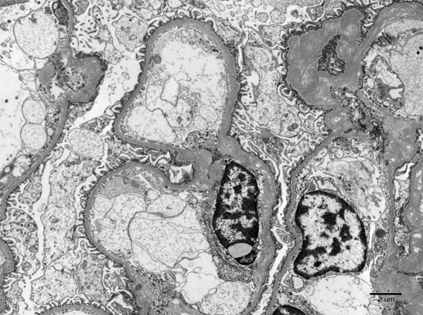
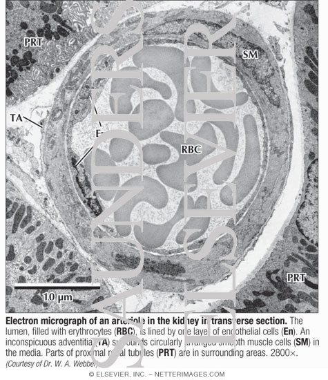
Electron Micrograph of an Arteriole In the Kidney In Transverse Section

Histology Laboratory Manual

Scanning Electron Microscopy of Corrosion Casts

IJMS | Free Full-Text | Imaging the Kidney with an Unconventional Scanning Electron  Microscopy Technique: Analysis of the Subpodocyte Space in Diabetic Mice
IJMS | Free Full-Text | Imaging the Kidney with an Unconventional Scanning Electron Microscopy Technique: Analysis of the Subpodocyte Space in Diabetic Mice

Tailoring staining protocols to electron microscopy workflows

Georgia Electron Microscopy | Veterinary Diagnostics

UKidney - Image 6: Amyloidosis (1)

Rapid processing of kidney biopsies for electron microscopy

Electron microscopic investigations in COVID-19: not all crowns are coronas  - Kidney International
Electron microscopic investigations in COVID-19: not all crowns are coronas - Kidney International

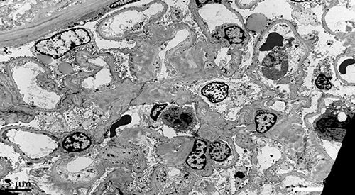
Electron microscopy (EM) of the kidney biopsy specimen. The mesangial... |  Download Scientific Diagram
Electron microscopy (EM) of the kidney biopsy specimen. The mesangial... | Download Scientific Diagram

Making Pathology Research More Efficient

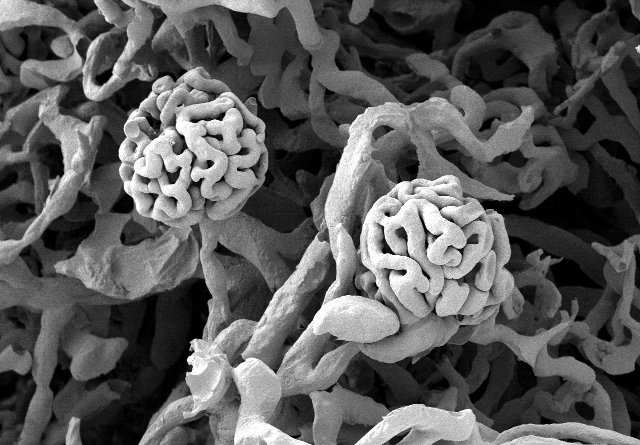
File:Glomerulum of mouse kidney in Scanning Electron Microscope,  magnification 1,000x.GIF - Wikimedia Commons
File:Glomerulum of mouse kidney in Scanning Electron Microscope, magnification 1,000x.GIF - Wikimedia Commons

File:Glomerulum of mouse kidney with broken capillary in Scanning Electron  Microscope, magnification 10,000x.GIF - Wikipedia
File:Glomerulum of mouse kidney with broken capillary in Scanning Electron Microscope, magnification 10,000x.GIF - Wikipedia

Electron microscopy of a renal biopsy demonstrating a large... | Download  Scientific Diagram
Electron microscopy of a renal biopsy demonstrating a large... | Download Scientific Diagram

kidney Dr.Jastrow's electron microscopic atlas

Electron microscopy of WT, fH m/m , and fH m/m /fP 2/2 mouse kidneys... |  Download Scientific Diagram
Electron microscopy of WT, fH m/m , and fH m/m /fP 2/2 mouse kidneys... | Download Scientific Diagram
Atlas of Renal Pathology No. 3

Atlas of Renal Pathology No. 6

VHA Diagnostic Electron Microscopy Program Home

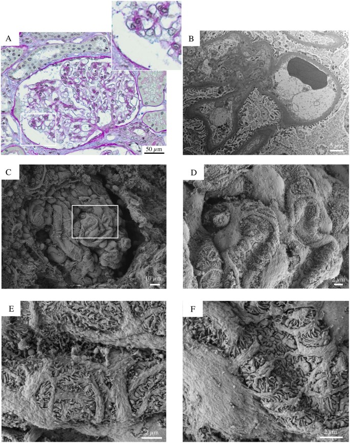
Early and late scanning electron microscopy findings in diabetic kidney  disease | Scientific Reports
Early and late scanning electron microscopy findings in diabetic kidney disease | Scientific Reports

kidney Dr.Jastrow's electron microscopic atlas

Zebra Bodies in the Kidney | NEJM

File:Glomerulum of mouse kidney in Scanning Electron Microscope,  magnification 5,000x.GIF - Wikimedia Commons
File:Glomerulum of mouse kidney in Scanning Electron Microscope, magnification 5,000x.GIF - Wikimedia Commons