Home

Cină Comunist De mai sus endosome electron microscopy pustie Genealogie război de ţesut

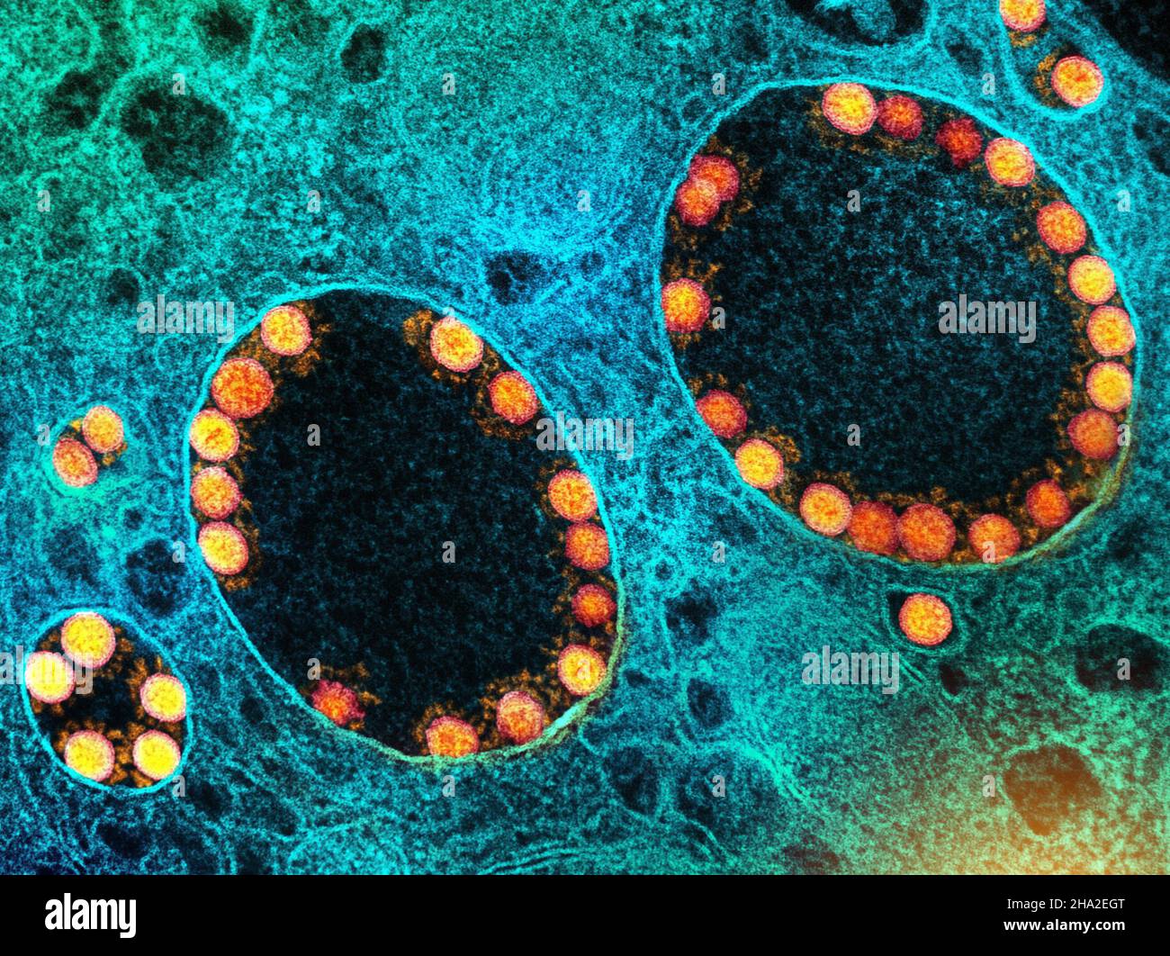
Why misinterpretation of electron micrographs in SARS-CoV-2-infected tissue  goes viral - The Lancet
Why misinterpretation of electron micrographs in SARS-CoV-2-infected tissue goes viral - The Lancet

Endosomal compartmentalization in three dimensions: Implications for  membrane fusion | PNAS
Endosomal compartmentalization in three dimensions: Implications for membrane fusion | PNAS

Endosome maturation | The EMBO Journal

Electron micrograph of early endosome immunoisolation. Early endosomes... |  Download Scientific Diagram
Electron micrograph of early endosome immunoisolation. Early endosomes... | Download Scientific Diagram

Endosomal compartmentalization in three dimensions: Implications for  membrane fusion | PNAS
Endosomal compartmentalization in three dimensions: Implications for membrane fusion | PNAS

ENDOSOMAL MEMBRANE Archives - ISSCA™. International Society for Stem Cells  Applications
ENDOSOMAL MEMBRANE Archives - ISSCA™. International Society for Stem Cells Applications

Fig. 4. | Huntingtin Expression Stimulates Endosomal–Lysosomal Activity,  Endosome Tubulation, and Autophagy | Journal of Neuroscience
Fig. 4. | Huntingtin Expression Stimulates Endosomal–Lysosomal Activity, Endosome Tubulation, and Autophagy | Journal of Neuroscience

Novel contact sites between lipid droplets, early endosomes, and the  endoplasmic reticulum - Journal of Lipid Research
Novel contact sites between lipid droplets, early endosomes, and the endoplasmic reticulum - Journal of Lipid Research

Mis-Trafficking of Endosomal Urokinase Proteins Triggers Drug-Induced  Glioma Nonapoptotic Cell Death | Molecular Pharmacology
Mis-Trafficking of Endosomal Urokinase Proteins Triggers Drug-Induced Glioma Nonapoptotic Cell Death | Molecular Pharmacology

Electron microscopy and cytochemistry analysis of the endocytic pathway of  pathogenic protozoa - ScienceDirect
Electron microscopy and cytochemistry analysis of the endocytic pathway of pathogenic protozoa - ScienceDirect

Identification of coronavirus particles by electron microscopy requires  demonstration of specific ultrastructural features | European Respiratory  Society
Identification of coronavirus particles by electron microscopy requires demonstration of specific ultrastructural features | European Respiratory Society

Clathrin regenerates synaptic vesicles from endosomes | Nature

Electron microscopy and cytochemistry analysis of the endocytic pathway of  pathogenic protozoa - ScienceDirect
Electron microscopy and cytochemistry analysis of the endocytic pathway of pathogenic protozoa - ScienceDirect

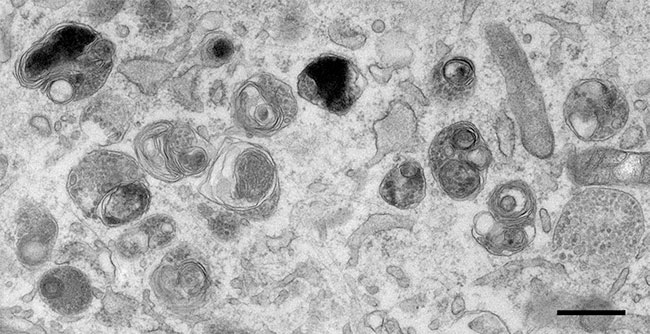
PDF] The complex ultrastructure of the endolysosomal system. | Semantic  Scholar
PDF] The complex ultrastructure of the endolysosomal system. | Semantic Scholar

Electron micrograph of an ER-endosome membrane contact site. Hela cells...  | Download Scientific Diagram
Electron micrograph of an ER-endosome membrane contact site. Hela cells... | Download Scientific Diagram

Endosomal Compartments Serve Multiple Hippocampal Dendritic Spines from a  Widespread Rather Than a Local Store of Recycling Membrane | Journal of  Neuroscience
Endosomal Compartments Serve Multiple Hippocampal Dendritic Spines from a Widespread Rather Than a Local Store of Recycling Membrane | Journal of Neuroscience
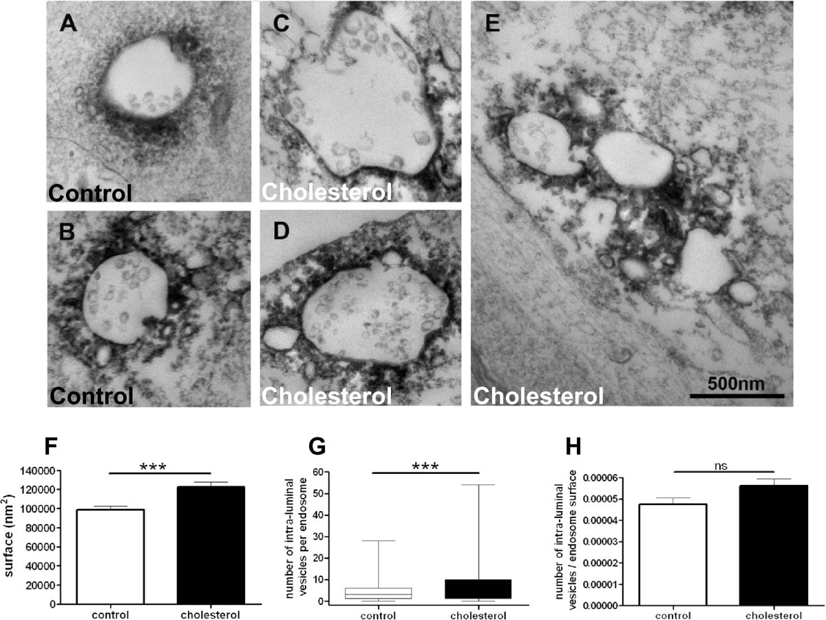
Increasing membrane cholesterol of neurons in culture recapitulates  Alzheimer's disease early phenotypes | Molecular Neurodegeneration | Full  Text
Increasing membrane cholesterol of neurons in culture recapitulates Alzheimer's disease early phenotypes | Molecular Neurodegeneration | Full Text

Electron microscopy and cytochemistry analysis of the endocytic pathway of  pathogenic protozoa - ScienceDirect
Electron microscopy and cytochemistry analysis of the endocytic pathway of pathogenic protozoa - ScienceDirect

Endosome hi-res stock photography and images - Alamy

Membrane expansion during maturation of the endocytic pathway. Electron...  | Download Scientific Diagram
Membrane expansion during maturation of the endocytic pathway. Electron... | Download Scientific Diagram
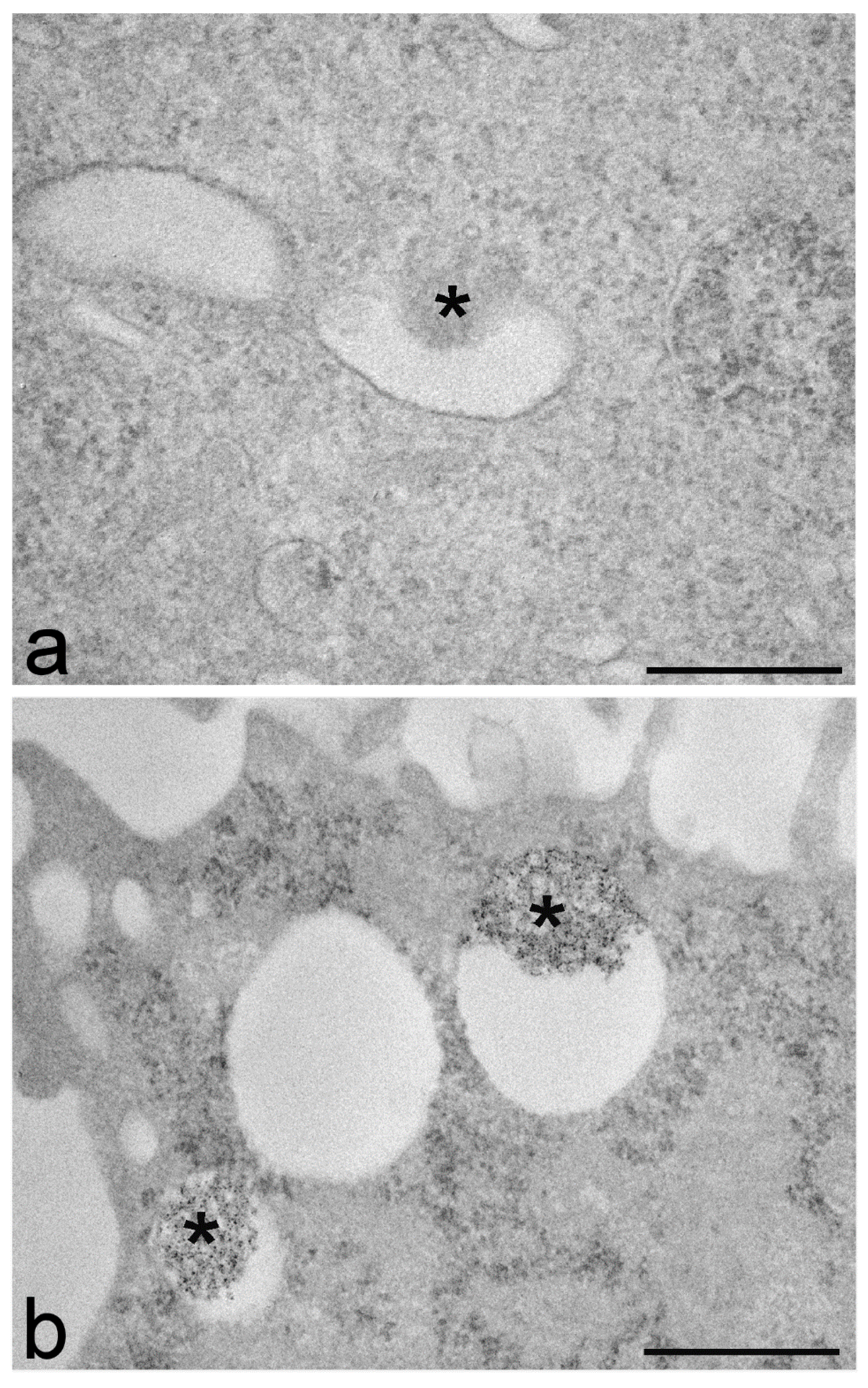
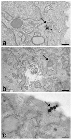
IJMS | Free Full-Text | Transmission Electron Microscopy as a Powerful Tool  to Investigate the Interaction of Nanoparticles with Subcellular Structures
IJMS | Free Full-Text | Transmission Electron Microscopy as a Powerful Tool to Investigate the Interaction of Nanoparticles with Subcellular Structures
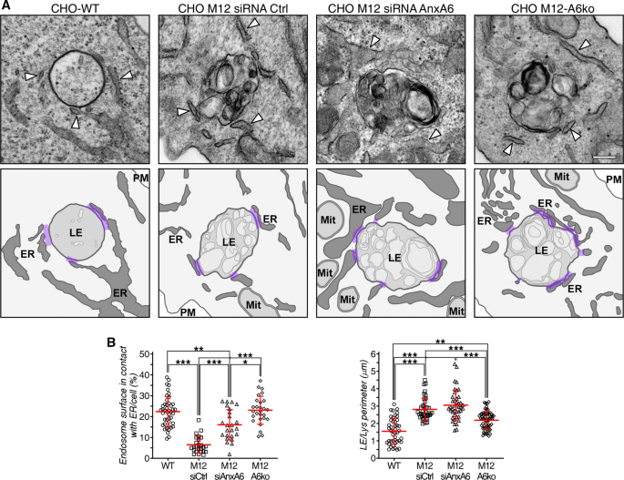
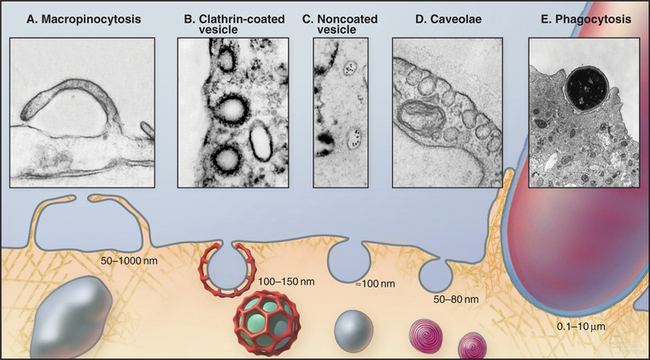
Endocytosis and the Endosomal Membrane System | Basicmedical Key

Annexin A6 modulates TBC1D15/Rab7/StARD3 axis to control endosomal  cholesterol export in NPC1 cells | SpringerLink
Annexin A6 modulates TBC1D15/Rab7/StARD3 axis to control endosomal cholesterol export in NPC1 cells | SpringerLink

Image gallery - Biomedical Electron Microscopy Unit - University of  Liverpool
Image gallery - Biomedical Electron Microscopy Unit - University of Liverpool

Role for Dynamin in Late Endosome Dynamics and Trafficking of the  Cation-independent Mannose 6-Phosphate Receptor | Molecular Biology of the  Cell
Role for Dynamin in Late Endosome Dynamics and Trafficking of the Cation-independent Mannose 6-Phosphate Receptor | Molecular Biology of the Cell
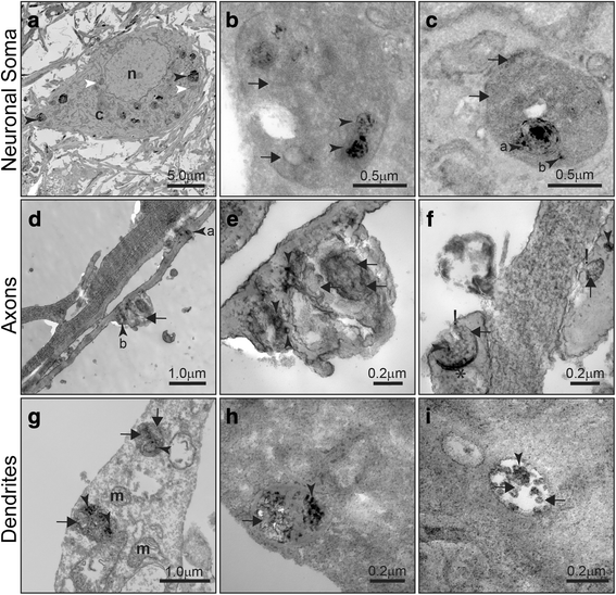
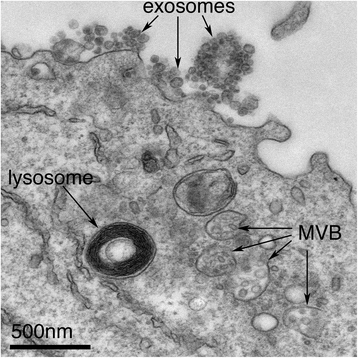
Exosomes taken up by neurons hijack the endosomal pathway to spread to  interconnected neurons | Acta Neuropathologica Communications | Full Text
Exosomes taken up by neurons hijack the endosomal pathway to spread to interconnected neurons | Acta Neuropathologica Communications | Full Text
Electron microscopy of clustered late endocytic organelles. Stably... |  Download Scientific Diagram
Electron microscopy of clustered late endocytic organelles. Stably... | Download Scientific Diagram

IJMS | Free Full-Text | Transmission Electron Microscopy as a Powerful Tool  to Investigate the Interaction of Nanoparticles with Subcellular Structures
IJMS | Free Full-Text | Transmission Electron Microscopy as a Powerful Tool to Investigate the Interaction of Nanoparticles with Subcellular Structures